




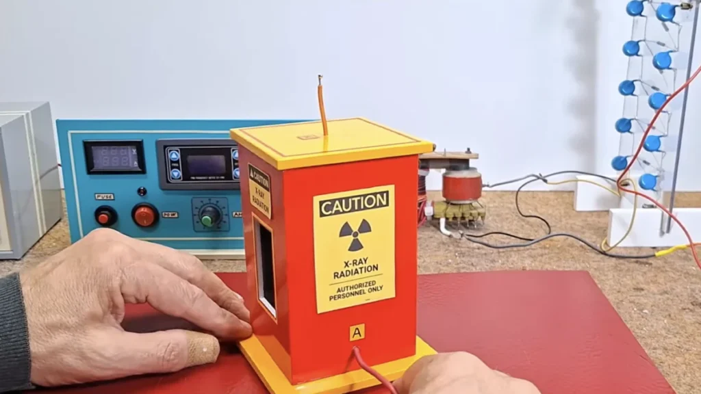
The intersection of amateur electronics and high-level physics has recently been highlighted by the work of Mirko Pavleski, a popular DIY enthusiast who successfully engineered a functional X-ray generator within a home workshop environment. While medical-grade imaging is traditionally the exclusive domain of licensed professionals and multi-million dollar facilities, Pavleski’s project demonstrates the underlying simplicity of the technology, provided one possesses the technical acumen to manipulate high-voltage currents and vacuum physics. The centerpiece of this experimental apparatus is a repurposed DY86 vacuum tube, a component once commonly found in vintage cathode-ray tube (CRT) television sets, repurposed here to serve as a rudimentary radiation source.
The achievement serves as both a historical tribute to the pioneers of radiology and a cautionary tale regarding the inherent dangers of ionizing radiation and high-voltage engineering. By utilizing discarded electronic components to replicate a Nobel Prize-winning discovery, the project underscores the accessibility of scientific principles while simultaneously raising important questions about safety and the regulation of amateur high-energy experiments.
The Historical Context of X-ray Discovery
To understand the significance of Pavleski’s DIY machine, one must look back to the late 19th century. In 1895, German physics professor Wilhelm Röntgen was experimenting with Crookes tubes—early experimental electrical discharge tubes—when he noticed a fluorescent effect on a nearby cardboard screen coated with barium platinocyanide. This occurred even when the tube was shielded by heavy black paper. Röntgen realized he had discovered a new form of electromagnetic radiation, which he dubbed "X-rays" due to their unknown nature.
Röntgen’s discovery revolutionized medicine almost overnight. By 1901, he was awarded the inaugural Nobel Prize in Physics. The fundamental mechanics of his original experiments remain the basis for modern radiography: high-velocity electrons are accelerated toward a metal target within a vacuum; upon impact, the kinetic energy of the electrons is converted into electromagnetic radiation in the X-ray spectrum. Pavleski’s use of a vacuum tube from an old television set effectively mimics this process, as these tubes were designed to handle high voltages and direct electron flow, making them accidental candidates for X-ray production when pushed beyond their original design parameters.
Technical Architecture and the Power Supply Chain
The primary challenge in building a DIY X-ray machine lies not in the tube itself, but in the generation of the massive electrical potential required to accelerate electrons to a speed sufficient to produce X-rays. For a vacuum tube like the DY86 to emit ionizing radiation, it must be subjected to voltages significantly higher than those found in standard household appliances.
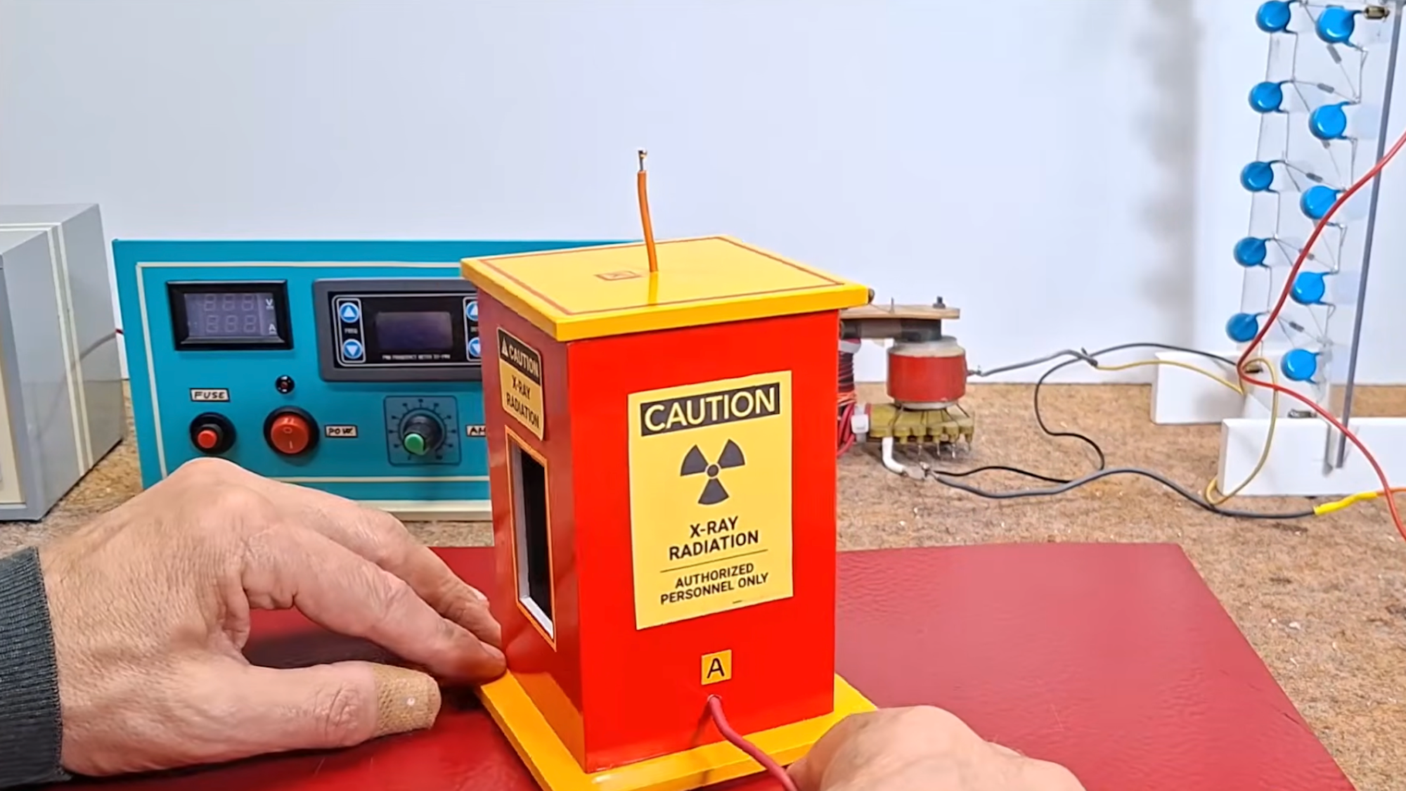
Pavleski engineered a complex power supply chain to achieve the necessary electrical environment. The process begins with a low-voltage DC generator, which provides the initial power source. This current is then fed into a high-voltage AC inverter, which converts the steady DC flow into an alternating current that can be manipulated by transformers. The next stage involves a specialized transformer that steps the voltage up to several thousand volts.
The final and most critical component in the power train is a voltage multiplier, often referred to as a Cockcroft-Walton generator. This circuit uses a ladder of capacitors and diodes to progressively "pump" the voltage to levels exceeding 30,000 to 50,000 volts. It is this extreme electrical pressure that forces electrons within the DY86 vacuum tube to strike the internal anode with enough force to generate "parasitic" X-rays.
The Trial and Error of High-Voltage Engineering
The path to a functional machine was fraught with technical setbacks, highlighting the volatility of the components involved. During the assembly and testing phases, Pavleski reportedly experienced the catastrophic failure of three separate vacuum tubes. These "explosions" are typically the result of thermal stress or internal arcing. When a vacuum tube designed for a specific voltage is subjected to the extreme pressures required for X-ray generation, the glass envelope can fail, or the internal metal components can overheat and vaporize, leading to a loss of vacuum and an immediate structural collapse.
Furthermore, the insulation requirements for such a device are immense. At 30,000 volts, electricity can "leak" through the air in the form of corona discharges or leap across gaps in the form of arcs. Ensuring that the high voltage remained contained within the circuit and directed solely toward the vacuum tube required precise component placement and the use of high-dielectric insulating materials.
The Imaging Process: From Radiation to Film
Once the apparatus was stabilized and producing a consistent beam of X-rays, Pavleski moved to the imaging phase. Unlike modern medical X-ray machines that use digital sensors (CMOS or CCD) to capture images instantly, this DIY project utilized traditional analog methods. Specifically, Pavleski used dental X-ray film—a small, high-resolution film packet typically used by dentists to check for cavities.
The process of capturing an image involves placing the subject—in this case, small electronic components like transistors and microSD cards—directly between the X-ray source and the film packet. When the machine is activated, the X-rays pass through the subject. Denser materials, such as the silicon and metal wiring inside a microSD card, absorb more radiation than the surrounding plastic. This creates a "shadow" on the film.
After exposure, the film must be developed using a chemical process. This involves a sequence of developer solution, a stop bath (often water), and a fixer solution. In Pavleski’s experiment, this manual development process took approximately 30 minutes per image. The resulting radiographs provided a clear, detailed look at the internal architecture of the workshop items, proving that the DIY machine was indeed producing ionizing radiation in the X-ray spectrum.
Safety Protocols and the Dangers of Ionizing Radiation
The most critical aspect of this project is the inherent danger involved. X-rays are a form of ionizing radiation, meaning they possess enough energy to detach electrons from atoms or molecules, which can cause significant damage to biological tissue and DNA. Prolonged or unprotected exposure to even low levels of X-rays can increase the risk of cancer, while high-dose exposure can cause immediate radiation burns or "deterministic effects."
In his demonstration, Pavleski emphasized the importance of safety by strictly limiting the subjects of his X-rays to inanimate objects. At no point were human body parts placed in the path of the beam. This is a vital distinction, as professional medical X-ray machines are equipped with precise lead collimators to narrow the beam, aluminum filters to remove low-energy "soft" X-rays that only add to skin dose without helping the image, and lead shielding to protect the operator.
Amateur setups often lack these sophisticated safety features. Furthermore, the DY86 tube used in the project is not shielded, meaning it emits X-rays in a wide pattern rather than a focused beam. Any person in the vicinity of such a device while it is operational would be at risk of secondary radiation exposure.

Regulatory and Ethical Implications
The construction of such devices falls into a complex regulatory gray area. In many jurisdictions, the possession of a radiation-producing device is strictly controlled by government agencies, such as the Nuclear Regulatory Commission (NRC) in the United States or the Environment Agency in the UK. While owning a vintage television is legal, modifying its components to intentionally produce ionizing radiation may violate health and safety statutes.
The rise of "Maker Culture" on platforms like YouTube has brought scientific experimentation into the home, but it also presents a challenge for public safety. Experts in the field of radiology often warn that while these projects are educational, they should not be replicated by those without professional training in radiation safety. The invisibility of X-rays makes them particularly dangerous; an experimenter could be receiving a significant dose of radiation without feeling any immediate sensation, only realizing the damage days or weeks later when symptoms of radiation sickness or skin lesions appear.
Broader Impact on Scientific Literacy
Despite the risks, Pavleski’s project has been lauded in some circles for its contribution to scientific literacy. By deconstructing the "black box" of medical technology, he demonstrates that the laws of physics are universal and accessible. The project encourages a deeper understanding of how the world works, moving beyond the consumer-level interface of modern technology to the fundamental electronic components that make it possible.
The use of "e-waste" or vintage components also highlights a growing movement toward the "Right to Repair" and the creative reuse of technology. In an era of planned obsolescence, Pavleski’s ability to find a new, albeit unintended, purpose for a decades-old vacuum tube serves as a powerful example of engineering resourcefulness.
Conclusion: A Bridge Between Eras
Mirko Pavleski’s DIY X-ray machine stands as a fascinating bridge between the experimental physics of the 1890s and the hobbyist engineering of the 2020s. It serves as a reminder that the foundational discoveries of modern science were often made with equipment that, by today’s standards, appears remarkably simple.
However, the transition of this technology from a controlled laboratory to a home workshop necessitates a high degree of responsibility. While the internal circuitry of a microSD card revealed by a repurposed TV tube is a triumph of amateur ingenuity, the invisible beams of radiation used to capture that image remain as potent and dangerous as they were when Wilhelm Röntgen first observed them over a century ago. The project serves its best purpose as an educational demonstration—a "look but don’t touch" exhibit in the vast gallery of modern digital science.