



Why do some new skills seem to click almost instantly while others remain frustratingly difficult even after repeated practice? For decades, neuroscientists have attributed the disparity in learning outcomes to a complex interplay of cognitive talent, deliberate practice, and synaptic plasticity. However, recent research suggests that the secret to mastering a new ability may lie not just in the brain’s architecture, but in the physiological state of the entire body. A landmark study conducted at Tohoku University, published on August 25, 2026, in the journal iScience, provides compelling evidence that the vagus nerve—the body’s primary communication highway—acts as a crucial regulator of long-term motor skill consolidation.
The Physiology of the Vagus Nerve
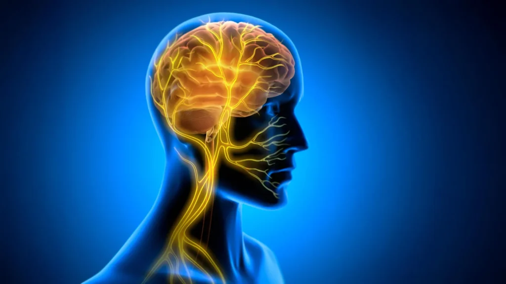
The vagus nerve is the longest cranial nerve in the human body, extending from the brainstem through the neck and down into the chest and abdomen. It functions as a bidirectional information superhighway, relaying sensory data from internal organs—such as the heart, lungs, and digestive tract—to the central nervous system, while simultaneously carrying regulatory commands from the brain to these organs.
For years, Vagus Nerve Stimulation (VNS) has been an established clinical intervention. Since its initial approval by the U.S. Food and Drug Administration in 1997 for treatment-resistant epilepsy, VNS has been refined for use in cases of severe depression and inflammatory disorders. Historically, the therapeutic success of VNS was attributed to its ability to modulate neurotransmitter systems, such as norepinephrine and acetylcholine, which are known to influence mood, arousal, and attention. However, the Tohoku University research team, led by Professor Ko Matsui, has identified a previously overlooked mechanism: the regulation of rhythmic blood volume fluctuations within the brain.
Chronology of the Tohoku Study
The investigation into this phenomenon began with a hypothesis: if the brain’s metabolic environment is influenced by peripheral signals, then targeted stimulation of the vagus nerve might be able to "prime" the brain to solidify memory after a training session.
To test this, researchers developed a micro-cuff electrode capable of interfacing with the left cervical vagus nerve in mice. The study followed a rigorous timeline:
- Initial Training Phase: Mice were introduced to a horizontal optokinetic response (HOKR) task. This cerebellum-dependent task requires the subject to track moving visual stripes, mirroring the natural eye movements humans exhibit when observing a passing train from a stationary platform.
- The Stimulation Protocol: Unlike previous studies that applied stimulation during the learning process, the Tohoku team introduced VNS exclusively after each training session.
- Observation Period: Over the course of five days, researchers tracked the mice’s performance, measuring the durability of the learned eye-tracking behavior.
- Data Synthesis: By August 2026, the researchers analyzed the correlation between the timing of stimulation, the resulting vascular rhythms in the cerebellum, and the final performance metrics of the mice.
The Discovery: Post-Practice Consolidation
One of the most striking findings of the study was the temporal specificity of the intervention. When VNS was administered during the learning task, it yielded negligible results. However, when applied immediately following the practice, the stimulation acted as a catalyst for long-term retention.
"The key point is that VNS was delivered only after training," explains Professor Matsui. "Our findings suggest that VNS may open a hidden window of opportunity for enhanced learning by making the brain environment more receptive to long-lasting change." This indicates that the brain requires a "consolidation phase" where the physical structure of neural pathways is hardened, and that external signals from the vagus nerve can optimize this period.
Rhythmic Vascular Oscillations
The study utilized advanced fiber photometry to observe the internal mechanics of the cerebellum, specifically the cerebellar flocculus. The data revealed a two-phase vascular response triggered by VNS: an initial, brief decrease in local blood volume followed by a sustained increase.
When the stimulation was repeated, these fluctuations stabilized into rhythmic oscillations. Remarkably, there was a direct positive correlation between the amplitude of these blood volume oscillations and the learning success observed by Day 5. This suggests that the body does not just signal the brain via chemical messengers; it modulates the brain’s metabolic supply chain through controlled vascular movement. "Our brains may be more strongly influenced by the body than we imagine," says lead author Junyu Chen. "By tuning the brain’s metabolic environment, including rhythmic vascular movements, we may eventually unlock capacities that would otherwise remain latent."
Broader Implications for Neuro-Rehabilitation
The implications of these findings extend far beyond the laboratory. If human brains respond to post-training VNS with similar vascular shifts, the potential for clinical applications is immense.
In the context of stroke rehabilitation, patients often struggle to re-learn motor skills such as walking or grasping objects. Current physical therapy relies on repetitive motion to induce neuroplasticity. By integrating VNS into a post-therapy regimen, clinicians might be able to accelerate the "closing" of the memory consolidation window, allowing patients to regain function more rapidly and durably.
Furthermore, the study challenges the traditional "brain-centric" view of human intelligence and skill acquisition. It reinforces the concept of the "embodied brain," where cognitive processes are inextricably linked to the physiological state of the organs. This shift in perspective could influence everything from educational techniques for neurodivergent students to advanced training protocols for elite athletes and musicians, who are constantly seeking methods to optimize their learning curves.
Limitations and Future Research
While the findings are groundbreaking, the research team is cautious about immediate human application. The current study was performed on mice, and the translation of vascular oscillation patterns from rodent models to human neurophysiology requires extensive clinical validation.
"Future research will focus on refining stimulation protocols and determining more precisely how communication between the brain and body supports long-term plasticity," the team noted in their concluding report. Scientists are particularly interested in determining whether the intensity, frequency, and duration of the stimulation can be tuned to maximize different types of learning, such as spatial memory versus linguistic acquisition.
There is also the question of safety and side effects. Because the vagus nerve innervates numerous critical organs, any therapeutic application in humans must ensure that the stimulation is localized enough to affect brain plasticity without causing adverse reactions in the digestive or cardiovascular systems.
A New Frontier in Neuroscience
The Tohoku University study serves as a critical junction in neuroscience, bridging the gap between visceral physiology and cognitive science. By identifying the vagus nerve as a lever for controlling the brain’s "receptivity," the researchers have provided a roadmap for potential future therapies that treat the human system as a unified whole.
As research continues, the scientific community expects to see a greater focus on "peripheral-to-central" pathways. If we can master the ability to send signals from the body to the brain to optimize learning, we may be on the cusp of a revolution in how we acquire new skills, recover from injury, and perhaps even enhance the natural cognitive limits of the human mind. The once-frustrating plateau of the learning curve may soon become a barrier that we can systematically overcome, provided we learn to speak the language of the body.