


This groundbreaking achievement represents a pivotal moment in neuroscience, as researchers from Duke-NUS Medical School and a consortium of international partners have successfully assembled one of the most complete single-cell maps of the developing human brain. The meticulously detailed atlas goes beyond mere structural representation, identifying nearly every cell type present during development, meticulously recording their unique genetic signatures, and illustrating the intricate ways these cells grow, differentiate, and interact within the complex neural architecture. Crucially, the research also provides a rigorous comparison of leading laboratory methods used to produce high-quality neurons, directly addressing critical challenges in the development of new therapeutic strategies for debilitating brain disorders such as Parkinson’s disease.
The Pressing Need for Precision in Parkinson’s Research
Parkinson’s disease stands as Singapore’s second most common neurodegenerative condition, affecting approximately three in every 1,000 individuals aged 50 and above. Globally, the prevalence is even more pronounced, with estimates suggesting that over 1% of the population above 60 years of age is afflicted, a figure projected to rise significantly with an aging global demographic. The economic burden of Parkinson’s is substantial, with healthcare costs in developed nations often running into billions of dollars annually, encompassing direct medical expenses, long-term care, and lost productivity. The disorder’s insidious progression specifically targets and harms midbrain dopaminergic neurons, which are vital for releasing dopamine, a neurotransmitter essential for regulating movement, motivation, and learning. The degeneration of these specific neurons leads to the characteristic motor symptoms of Parkinson’s, including tremors, bradykinesia (slowness of movement), rigidity, and postural instability, alongside a host of non-motor symptoms like cognitive impairment, depression, and sleep disturbances.
Current treatments for Parkinson’s disease primarily focus on symptomatic relief, with levodopa remaining the gold standard for managing motor symptoms. While effective initially, its long-term use is associated with debilitating side effects such as dyskinesias (involuntary movements). Other therapeutic avenues include deep brain stimulation (DBS) for advanced cases, but these interventions do not halt or reverse the underlying neurodegeneration. This landscape underscores the urgent need for disease-modifying therapies, and cell replacement therapy, which aims to restore lost dopaminergic neurons, has long been viewed as one of the most promising frontiers. However, translating this promise into effective clinical treatments has been fraught with challenges, largely due to the difficulty in reliably producing pure populations of functional dopaminergic neurons and ensuring their safe and effective integration into the patient’s brain without unwanted side effects.
BrainSTEM: A Novel Two-Step Mapping Approach
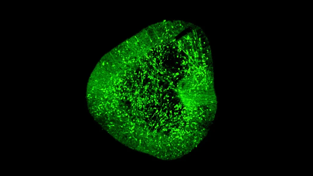
To address these critical hurdles and to bring clarity to the precise mechanisms by which dopaminergic neurons form and mature, particularly in laboratory-generated settings, the research team developed an innovative two-step mapping approach dubbed BrainSTEM (Brain Single-cell Two tiEr Mapping). This sophisticated methodology was crucial for navigating the immense cellular complexity of the developing brain. In a significant collaborative effort, involving key partners such as the University of Sydney, the researchers meticulously profiled nearly 680,000 individual cells harvested from the fetal brain. This extensive initial profiling enabled them to chart the entire cellular landscape of the developing human brain at an unprecedented level of detail, providing a foundational understanding of the diverse cell types and their developmental trajectories.
The second, higher-resolution projection within the BrainSTEM framework then specifically targeted the midbrain region with enhanced precision. This focused approach allowed the researchers to pinpoint and characterize dopaminergic neurons with exquisite accuracy, distinguishing them from other closely related but functionally distinct cell types. The resulting "comprehensive reference map" is not merely a descriptive tool; it has been meticulously designed to serve as a new global standard. This benchmark allows researchers worldwide to rigorously evaluate the accuracy and fidelity of laboratory-produced midbrain models against the true biological complexity of the human brain, ensuring that therapeutic cells are as authentic and effective as possible.
Unmasking Imperfections in Current Lab Practices
A critical finding emerging from the study, published in the esteemed journal Science Advances, revealed a significant limitation in existing laboratory protocols for generating neurons. The research unequivocally demonstrated that several methods commonly employed to grow midbrain cells in vitro also inadvertently generated unwanted cells originating from other brain regions. These "off-target" cell populations pose substantial risks in the context of cell replacement therapy. Their presence could lead to reduced therapeutic efficacy, as non-dopaminergic cells would not contribute to dopamine production, and more critically, they could introduce unpredictable side effects, including the formation of tumors or aberrant neural activity, jeopardizing patient safety.
These findings carry profound implications, indicating that both the experimental protocols used for cell differentiation and the subsequent data analysis pipelines require considerable refinement. The ability to detect and effectively remove such off-target populations is paramount for ensuring the purity, safety, and ultimate efficacy of any cell-based therapy destined for human application. BrainSTEM’s unparalleled precision offers the tools necessary to achieve this level of scrutiny, moving the field closer to generating truly transplant-ready cellular products.
The Evolution of Brain Mapping: From Macro to Micro
The journey to mapping the human brain has been a long and arduous one, evolving dramatically over centuries. Early efforts relied on macroscopic anatomical observations and later, rudimentary microscopy, providing a foundational understanding of brain structures. The 20th century saw the advent of more sophisticated staining techniques, like the Golgi stain, which allowed visualization of individual neurons, albeit sparsely. However, these methods offered limited insight into the molecular identities or functional states of different cell types.
The true revolution began with the sequencing of the human genome and the subsequent development of high-throughput molecular techniques. The past decade has witnessed a paradigm shift with the advent of single-cell sequencing technologies. Prior to this, researchers relied on "bulk" tissue analysis, which averaged out the molecular profiles of millions of cells, obscuring the vast cellular heterogeneity within a given brain region. Single-cell RNA sequencing (scRNA-seq) has transformed this by allowing scientists to profile the gene expression of individual cells, revealing distinct cell types, states, and developmental trajectories within complex tissues.
BrainSTEM stands at the forefront of this revolution. While initiatives like the Human Brain Project and the BRAIN Initiative have aimed to map the entire brain, BrainSTEM’s unique contribution lies in its focus on developmental stages and its therapeutic application, specifically for Parkinson’s. By creating a reference map of unparalleled resolution for dopaminergic neuron development, it provides a critical benchmark that previous, broader atlases could not offer with the same level of granularity and functional specificity required for cell therapy. This chronological progression from broad structural mapping to intricate single-cell molecular profiling underscores the increasing precision that defines contemporary neuroscience.
Expert Perspectives on BrainSTEM’s Impact
The researchers involved in the study articulated the profound implications of their work. Dr. Hilary Toh, an MD-PhD candidate from the Neuroscience & Behavioural Disorders program at Duke-NUS Medical School and one of the paper’s first authors, emphasized the practical utility of the atlas. "Our data-driven blueprint helps scientists produce high-yield midbrain dopaminergic neurons that faithfully reflect human biology," Dr. Toh stated. "Grafts of this quality are pivotal to increasing cell therapy efficacy and minimizing side effects, paving the way to offer alternative therapies to people living with Parkinson’s disease." Her statement underscores the direct translational potential, highlighting how enhanced cellular purity and authenticity are non-negotiable for successful clinical outcomes.
Dr. John Ouyang, Principal Research Scientist from Duke-NUS’ Centre for Computational Biology and a senior author of the study, elaborated on the technological prowess and future applications. "By mapping the brain at single-cell resolution, BrainSTEM gives us the precision to distinguish even subtle off-target cell populations," he explained. "This rich cellular detail provides a critical foundation for AI-driven models that will transform how we group patients and design targeted therapies for neurodegenerative diseases." Dr. Ouyang’s comments point towards the synergy between high-resolution biological data and artificial intelligence, envisioning a future where personalized medicine for neurological disorders is guided by deep cellular insights. The ability to identify minute differences in cell populations could allow AI algorithms to discern distinct disease subtypes within Parkinson’s, leading to more tailored and effective treatments.
Assistant Professor Alfred Sun from Duke-NUS’ Neuroscience & Behavioural Disorders programme, also a senior author of the paper, highlighted the transformative nature of BrainSTEM for the field of brain modeling. "BrainSTEM marks a significant step forward in brain modeling. By delivering a rigorous, data-driven approach, it will speed the development of reliable cell therapies for Parkinson’s disease. We’re setting a new standard to ensure the next generation of Parkinson’s models truly reflects human biology." His remarks underscore the establishment of a new benchmark, suggesting that future research and therapeutic development will be held to a higher standard of biological fidelity and data rigor.
Professor Patrick Tan, Senior Vice-Dean for Research at Duke-NUS, provided a broader institutional perspective on the study’s significance. "This study redefines the benchmark — establishing multi-tier mapping as essential for capturing cellular detail in complex biological systems. By revealing how the human midbrain develops in such detail, we will accelerate Parkinson’s research and cell therapy, delivering better care and offer hope to people living with the disease." Professor Tan’s statement encapsulates the overarching impact, positioning multi-tier mapping as an indispensable methodology for unlocking the complexities of biological systems and accelerating the pace of translational research for tangible patient benefit.
Broader Implications and Future Trajectories
The impact of the BrainSTEM atlas extends far beyond the immediate advancements in Parkinson’s disease research. The researchers have committed to releasing their comprehensive brain atlases as open-source references and providing the multi-tier mapping approach as an "out-of-the-box package." This commitment to open science is transformative, democratizing access to cutting-edge tools and data for the global scientific community. Because BrainSTEM is designed to be broadly applicable for isolating and characterizing "any cell type in the brain," laboratories worldwide can leverage this robust framework to deepen insights into various neurological conditions, streamline their experimental workflows, and accelerate the pace of discovery across the entire spectrum of neuroscience. This collaborative ethos fosters rapid innovation, as researchers can build upon a shared, high-quality foundation.
The integration of such rich cellular detail with artificial intelligence platforms, as alluded to by Dr. Ouyang, holds immense promise. AI algorithms, fed with this granular data, could revolutionize how neurodegenerative diseases are classified, how patients are grouped for clinical trials, and how personalized therapeutic strategies are designed. For instance, AI could identify subtle molecular signatures that differentiate rapidly progressing Parkinson’s from slower forms, allowing for earlier and more aggressive intervention where needed.
Furthermore, the precise understanding of cellular development and interaction provided by BrainSTEM will significantly aid in drug discovery and development. By identifying critical developmental pathways and specific genetic signatures of healthy versus diseased cells, researchers can pinpoint novel therapeutic targets for small molecules or biologics. The ability to accurately assess the purity and identity of lab-grown neurons also de-risks preclinical development, reducing the likelihood of costly failures in later-stage clinical trials.
While the immediate focus is on Parkinson’s, the methodological flexibility of BrainSTEM means its utility can be extended to other devastating neurodegenerative conditions like Alzheimer’s disease, Huntington’s disease, and Amyotrophic Lateral Sclerosis (ALS), as well as various neurodevelopmental disorders. By providing a common, rigorous standard for cell identification and quality control, BrainSTEM has the potential to accelerate cell therapy development across a broader range of brain diseases. From a clinical translation perspective, this research offers a clear pathway to safer and more efficacious cell therapies, which could ultimately lead to improved quality of life for millions of patients and a reduction in the substantial healthcare burden associated with these chronic conditions.
This monumental work received vital support from several key programs, including the USyd-NUS Ignition Grant and the Duke-NUS Parkinson’s Research Fund, the latter generously supported by a donation from The Ida C. Morris Falk Foundation. Duke-NUS Medical School continues to solidify its position as a global leader in medical research and education, demonstrating an unwavering commitment to enhancing patient care through rigorous scientific innovation. The successful completion and publication of this study represent a significant advancement in ongoing international efforts to unravel the fundamental mechanisms governing brain function and development, laying a critical foundation for the creation of truly transformative therapeutic strategies for a wide array of neurological conditions.