



Why do some new skills seem to click almost instantly while others remain frustratingly difficult even after repeated practice? The difference may involve more than innate talent or dedicated effort; it may hinge on whether the physiological state of the brain is primed to translate transient practice into permanent neurological storage. A groundbreaking study published in the journal iScience on August 25, 2026, by researchers at Tohoku University, offers a compelling answer: the secret to lasting learning may reside in the body’s ability to communicate with the brain through the vagus nerve.
The study, which examined motor skill acquisition in murine models, suggests that stimulating the vagus nerve following a training session—rather than during it—can significantly strengthen the retention of new motor skills. By identifying a mechanism involving rhythmic vascular oscillations within the brain, the research team has opened a new frontier in neuroscience, suggesting that the brain’s "metabolic environment" is a critical, yet previously overlooked, component of memory consolidation.
The Anatomy of the Brain-Body Connection
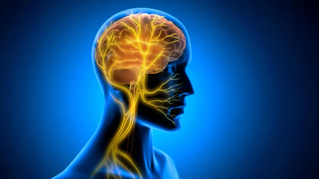
The vagus nerve is one of the most complex components of the human nervous system. Serving as the primary conduit for the parasympathetic nervous system, it acts as a bidirectional communication superhighway, transmitting sensory data from the heart, lungs, and digestive tract to the brainstem, while simultaneously carrying regulatory signals from the brain back to these organs.
For decades, the medical community has utilized Vagus Nerve Stimulation (VNS) as a therapeutic intervention for treatment-resistant epilepsy and major depressive disorder. Traditionally, VNS has been viewed through the lens of neurotransmitter modulation—the idea that electrical pulses influence the release of chemicals like norepinephrine or acetylcholine. However, this new research from Tohoku University posits a secondary, purely physical mechanism: the regulation of intracranial blood volume.
Chronology of the Discovery
The research, led by Professor Ko Matsui and lead author Junyu Chen, unfolded through a systematic investigation into the cerebellum, the region of the brain responsible for fine-tuning movement and motor learning.
The experiment was conducted in several distinct phases:
- Preparation (Early 2026): Researchers developed a specialized cuff electrode, a micro-scale device designed to maintain constant contact with the left cervical vagus nerve in mice without hindering their natural behavior.
- Training Phase: The subjects were trained in the horizontal optokinetic response (HOKR). This task requires the subject to track moving visual stripes, an automatic reflex similar to how a passenger on a train watches the landscape shift outside a window.
- Intervention (August 2026): Unlike previous studies that applied stimulation concurrently with learning, this team isolated the VNS to the post-training window.
- Observation: Following the stimulation, the team utilized fiber photometry to monitor real-time changes in blood volume within the cerebellar flocculus—the specific area of the brain governing the HOKR reflex.
The results were stark. When VNS was applied after the training sessions, there was no immediate impact on performance. However, by the following day, the mice subjected to the stimulation demonstrated significantly higher retention rates than control groups. The findings indicate that VNS does not teach the skill; rather, it creates the biological conditions necessary for the brain to "lock in" the information provided during the training session.
The Role of Vascular Oscillations
Perhaps the most significant finding in the study is the discovery of "rhythmic vascular response." By observing blood volume near the cerebellar flocculus, the researchers noted that a single VNS pulse induced a two-phase vascular response: an initial, brief contraction, followed by a delayed expansion.
When these pulses were repeated, they created rhythmic oscillations in blood volume. The data suggested a direct correlation: mice that exhibited more pronounced rhythmic oscillations on the day of training consistently outperformed their peers by the fifth day of the experiment.
"Our findings suggest that VNS may open a hidden window of opportunity for enhanced learning by making the brain environment more receptive to long-lasting change," stated Professor Ko Matsui. This implies that the brain’s vascular system acts as a gatekeeper for memory. If the blood flow environment is properly "tuned," the neural pathways associated with a new skill can be stabilized more effectively.
Implications for Human Learning and Neurorehabilitation
The implications of this research for human neurology are profound. Currently, neurorehabilitation for stroke victims or those recovering from traumatic brain injuries relies heavily on repetitive physical therapy. The efficiency of this process is often limited by "plateaus," where patients struggle to retain the gains made during therapy sessions.
If VNS can be refined to optimize the metabolic environment of the human brain post-therapy, it could potentially accelerate recovery timelines. By utilizing the vagus nerve as a remote-control switch for brain plasticity, clinicians might be able to artificially extend the duration of the brain’s "receptive state."
"Our brains may be more strongly influenced by the body than we imagine," noted lead author Junyu Chen. "By tuning the brain’s metabolic environment, including rhythmic vascular movements, we may eventually unlock capacities that would otherwise remain latent."
Statistical Context and Scientific Peer Response
While the study is limited to murine models, the use of HOKR as a model for learning is highly regarded in the field of neuroscience. The HOKR is a cerebellum-dependent task, and the neural circuits involved are structurally similar to those in humans.
External experts in neurophysiology have noted that the study’s focus on the timing of the intervention—post-practice rather than mid-practice—addresses a common flaw in previous VNS research. By separating the acquisition phase from the consolidation phase, the researchers have provided a clearer view of the mechanics behind long-term potentiation (LTP).
Current data in the field of neuromodulation suggest that the market for VNS devices is expected to grow significantly as applications expand from epilepsy to cognitive enhancement and motor recovery. However, the Tohoku University team cautions that the transition from mouse to human will require years of rigorous clinical trials. The next steps for the team involve mapping the exact frequency and intensity of stimulation required to trigger these vascular rhythms without disrupting systemic blood pressure or cardiac function.
Future Research Directions
The research published in iScience is far from a finished product. The team is currently looking to answer three critical questions for future development:
- Dose-Response Relationship: What is the minimal effective dose of VNS required to stimulate the necessary vascular rhythms?
- Safety Profile: Does repeated, long-term stimulation of the vagus nerve have unintended consequences on the autonomic regulation of the heart and digestive system?
- Generality: Can this method be applied to non-motor learning, such as language acquisition or cognitive memory tasks, or is the benefit exclusive to the cerebellum?
As the medical community continues to explore the "brain-body" axis, this research serves as a reminder that the brain does not operate in a vacuum. It is a biological organ dependent on the metabolic rhythm of the entire body. By mastering the communication channels between the vagus nerve and the brain’s vascular system, science may soon be able to assist the human brain in doing what it does best: learning, storing, and mastering complex information with unprecedented efficiency.
The study highlights a shift in focus from simply treating brain pathology to actively managing the environment in which learning takes place. Whether this leads to new commercial therapies or a deeper understanding of human potential, the August 2026 findings represent a significant milestone in the quest to understand the mechanics of the mind.