

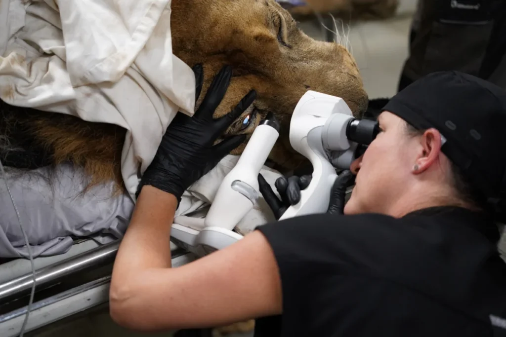
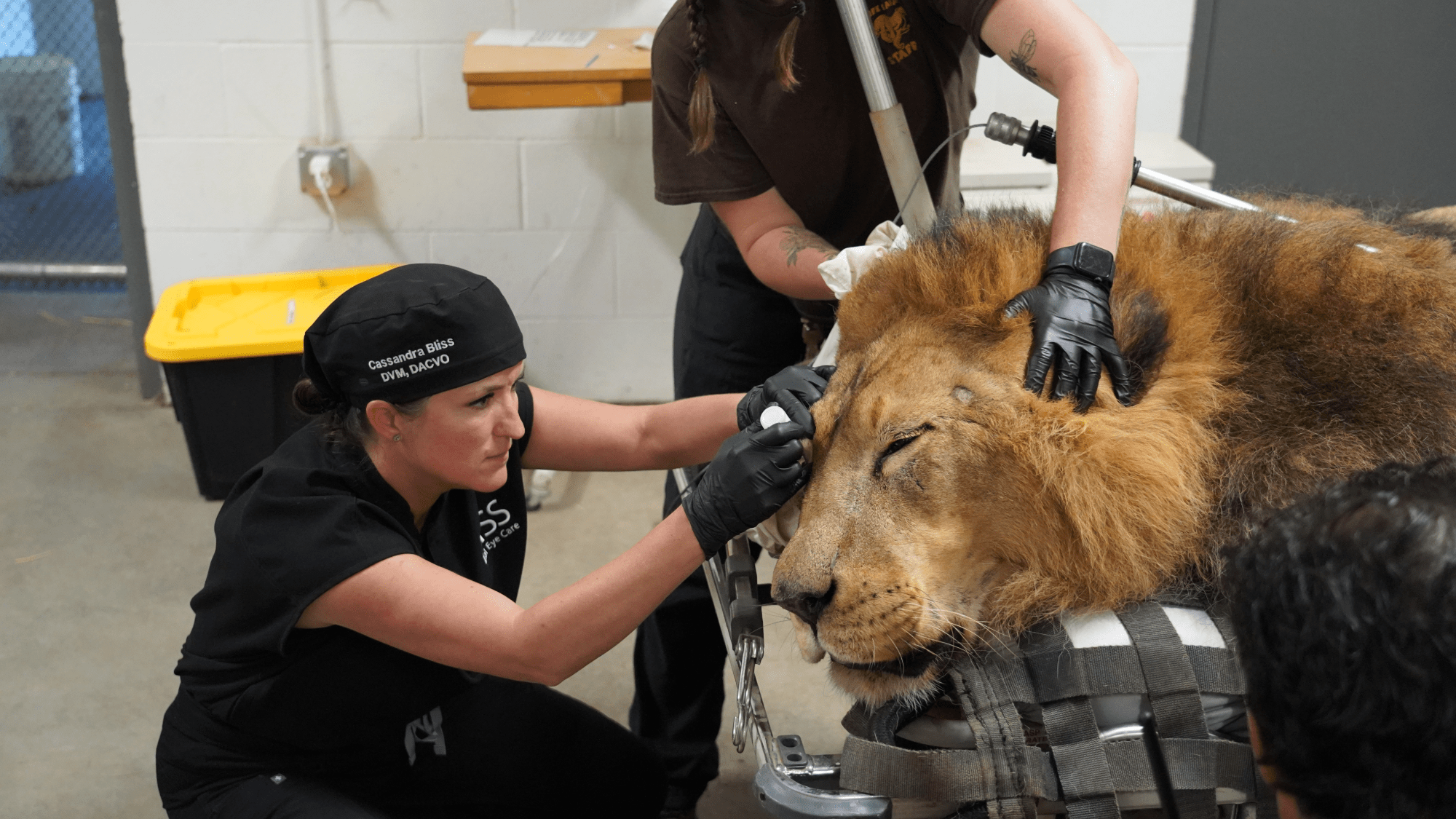
In a remarkable convergence of advanced medical technology and conservation expertise, a multi-disciplinary team of veterinary specialists successfully performed bilateral cataract surgery on an adult African lion named Tsavo at Wildlife Safari in Winston, Oregon. The procedure, conducted on June 12, marks a significant milestone in zoological medicine, restoring clear vision to a 400-pound predator through techniques that closely mirror those used in human ophthalmology. Tsavo, an 11-year-old lion who has resided at the wildlife park since 2015, had been suffering from progressive vision loss for over six months, a condition that severely impacted his ability to navigate his environment and interact with his pride. The success of the operation, which involved the implantation of custom-manufactured artificial lenses, highlights the growing capabilities of specialized veterinary care in improving the welfare of captive endangered species.
The Patient: From Orphaned Cub to Conservation Ambassador
Tsavo’s journey to the operating table began more than a decade ago. Rescued as an orphaned cub in Qatar in 2013, Tsavo was eventually relocated to the United States to live at Wildlife Safari, a 600-acre non-profit drive-through park dedicated to conservation and education. Over the years, Tsavo became a central figure in the park’s lion pride, contributing to the public’s understanding of the African lion (Panthera leo), a species currently classified as vulnerable by the International Union for Conservation of Nature (IUCN).
The first signs of Tsavo’s ocular decline were noted by the park’s carnivore care team in late 2025. Caretakers observed subtle changes in his behavior, including increased hesitancy when navigating uneven terrain and a diminished response to visual enrichment. A formal diagnostic exam confirmed the presence of bilateral cataracts—a condition characterized by the clouding of the eye’s natural lens. While cataracts are a frequent occurrence in aging domestic cats and humans, they present unique challenges in large felids due to the sheer scale of the anatomy and the complexities of administering general anesthesia to a high-risk apex predator.
Chronology of the Surgical Intervention
The decision to proceed with surgery was not made lightly. It required months of meticulous planning, international coordination, and the procurement of specialized equipment. The medical team was co-led by Dr. Cassandra Bliss of Bliss Animal Eye Care and Dr. Benjamin Alcantar, the Head Veterinarian at Wildlife Safari. This duo had previously collaborated on similar procedures, having successfully treated two other lions at the park for cataracts in the preceding year.
On the morning of June 12, the surgical suite was prepared with equipment sourced from global medical technology firms. The timeline of the procedure was as follows:
- 1:00 PM – Anesthesia Induction: The process began with the careful sedation of Tsavo in a controlled environment. Given the lion’s size and metabolic rate, the anesthesia team had to monitor his vitals with extreme precision to ensure stability throughout the procedure.
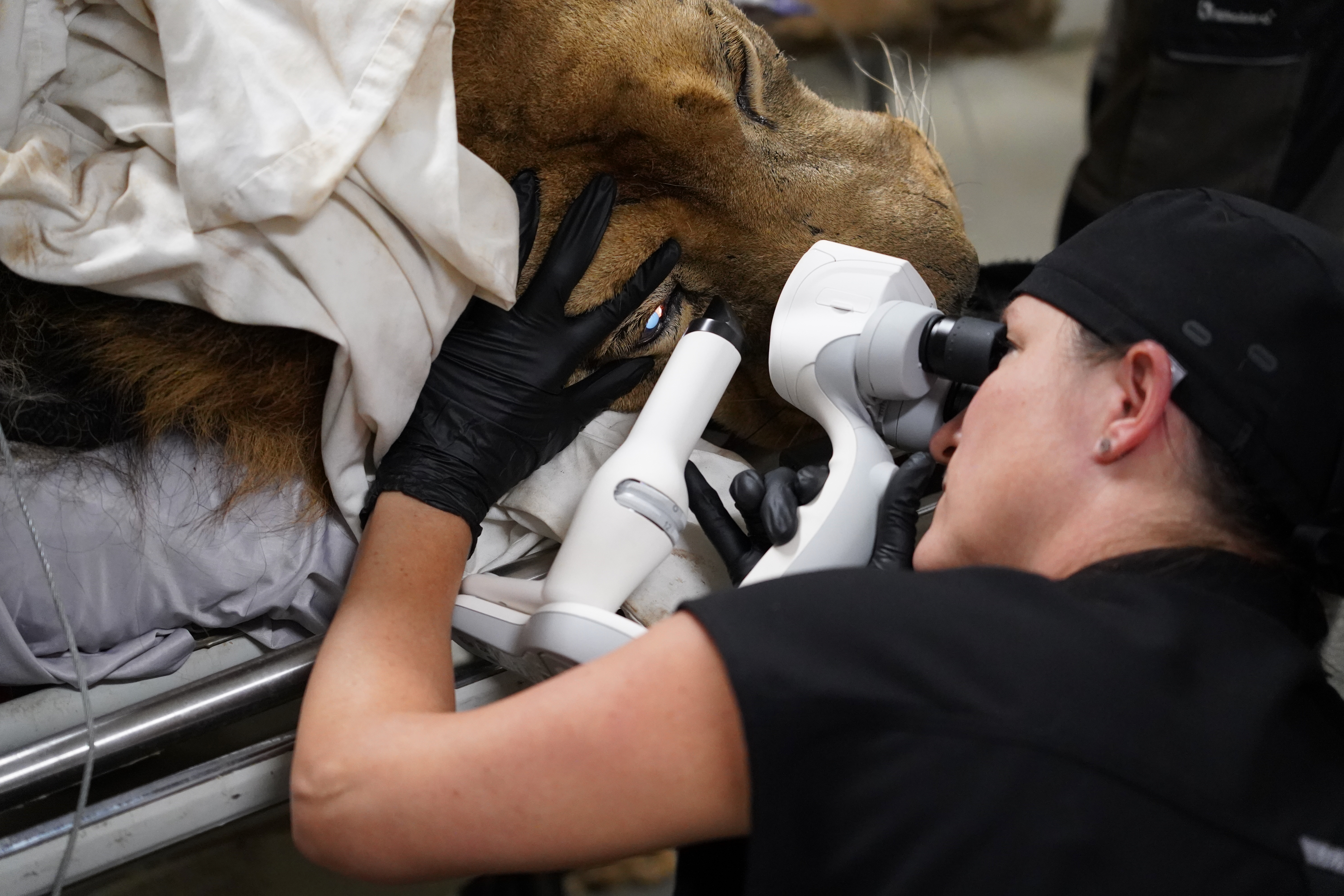
- 2:00 PM – Incision and Phacoemulsification: The surgery commenced. Using a technique known as phacoemulsification, the surgeons used ultrasonic vibrations to break up the clouded natural lenses. These fragments were then aspirated from the eye through a microscopic incision.
- 3:30 PM – Lens Implantation: Once the cataractous material was removed, the team inserted custom-made intraocular lenses (IOLs). These lenses were specifically engineered to match the refractive power and physical dimensions of a lion’s eye, which is significantly larger than that of a human.
- 4:45 PM – Completion and Suturing: The surgeons finalized the placement of the lenses and closed the incisions. The entire operation lasted less than three hours, a testament to the efficiency of the collaborative team.
- 5:15 PM – Recovery Initiation: Tsavo was transported back to the specialized "Lion Hut" recovery area. By late afternoon, he was beginning to wake from anesthesia under the continuous supervision of the carnivore team.
Comparative Anatomy and Technical Challenges
While the fundamental mechanics of cataract surgery are consistent across species, the scale of a lion’s eye introduces significant variables. A human eye typically has a diameter of about 24 millimeters, whereas a lion’s eye can exceed 36 to 40 millimeters. This size difference necessitates specialized surgical instruments and a higher volume of viscoelastics—clear, gel-like substances used to maintain the shape of the eye during surgery.
The artificial lenses used for Tsavo were the result of precise optical calculations. In humans, IOLs are standardized, but for a lion, the lenses must be robust enough to withstand the pressures of a predator’s ocular muscles while providing the specific focal length required for binocular vision. Dr. Bliss noted that the surgical process is "almost identical to modern human cataract surgery," yet the stakes are heightened by the patient’s nature. Unlike a human patient, a lion cannot be instructed to remain still or avoid rubbing its eyes post-operatively, requiring the medical team to utilize long-acting medications and specialized recovery enclosures.
The Role of Binocular Vision in Apex Predators
The restoration of Tsavo’s sight is more than a matter of visual clarity; it is a restoration of his biological identity. Lions are highly dependent on binocular vision and depth perception for their social and physical behaviors. In the wild, these traits are essential for hunting and territorial defense. In a managed environment like Wildlife Safari, depth perception is critical for safely navigating enclosures, jumping onto platforms, and accurately interpreting the body language of other lions in the pride.
"Vision is one of the primary ways lions interact with their environment," Dr. Bliss explained following the surgery. "Restoring sight isn’t just about seeing again; it’s about allowing an animal to navigate its world, engage with enrichment, recognize familiar caretakers and companions, and express the natural behaviors that contribute to its quality of life."
The loss of depth perception can lead to chronic stress in large carnivores, as they become unsure of their surroundings. By replacing the clouded lenses with clear artificial ones, the veterinary team has effectively "reset" Tsavo’s sensory input, allowing him to regain his confidence and status within the pride.
Collaborative Success and Global Expertise
The successful outcome for Tsavo was made possible by an international network of support. The equipment and lenses used in the procedure represent the pinnacle of current veterinary ophthalmology. This case serves as a prime example of "One Medicine"—the concept that human and animal health are linked and that medical advancements in one field can and should benefit the other.
Dr. Benjamin Alcantar emphasized that these procedures are part of a broader commitment to animal welfare at Wildlife Safari. The park has established itself as a leader in specialized veterinary care for big cats, utilizing its experiences to contribute to a global database of knowledge that helps other zoos and sanctuaries worldwide. The previous surgeries conducted by Bliss and Alcantar in 2024 provided the necessary data and "proof of concept" to ensure that Tsavo’s operation was as safe and predictable as possible.
Post-Operative Outlook and Broader Implications
As of the days following the surgery, Tsavo is reported to be recovering well. The carnivore team at Wildlife Safari is administering a regimen of anti-inflammatory and antibiotic eye drops, disguised in his food, to prevent infection and reduce swelling. Early observations suggest that Tsavo is already showing increased awareness of his surroundings, responding more quickly to visual cues from his keepers.
The implications of this surgery extend beyond one lion in Oregon. As captive populations of vulnerable species age, the demand for geriatric veterinary care—including ophthalmology, oncology, and orthopedics—is rising. Tsavo’s case demonstrates that even the most complex human medical interventions can be adapted for wildlife, provided there is sufficient collaboration between specialists and zoological institutions.
Furthermore, the success of such surgeries reinforces the role of modern wildlife parks as centers of medical excellence. While the primary goal of Wildlife Safari is conservation and education, the ability to provide state-of-the-art medical care is a vital component of that mission. By ensuring that ambassadors like Tsavo live long, healthy, and sighted lives, these institutions can continue to inspire the public to support the protection of African lions in the wild.
In the coming weeks, Tsavo will undergo several follow-up examinations to monitor the stability of the intraocular lenses and the healing of the corneal incisions. If his recovery continues on its current trajectory, he will soon be reunited with his pride, once again able to look out over the Oregon landscape with the clear, sharp gaze of a healthy African lion. This successful intervention stands as a testament to the dedication of the veterinary profession and the sophisticated technology now available to protect and care for the world’s most majestic creatures.