


A groundbreaking discovery by researchers at the Mark and Mary Stevens Neuroimaging and Informatics Institute (Stevens INI) at the Keck School of Medicine of USC has fundamentally altered our understanding of one of the brain’s most crucial regions for learning and memory. Published in the prestigious journal Nature Communications, their findings reveal a previously unrecognized organizational pattern within the CA1 section of a mouse’s hippocampus, identifying four distinct layers of specialized cell types. This revelation offers unprecedented insight into the intricate pathways of information processing within this vital brain structure and provides critical clues into the selective vulnerability of certain neuron types in debilitating neurological conditions such as Alzheimer’s disease and epilepsy.
For decades, the hippocampus has been a focal point of neuroscience research due to its indispensable role in forming new memories, facilitating spatial navigation, and modulating emotional responses. Its importance was famously underscored by the case of Patient H.M., whose severe anterograde amnesia following hippocampal removal highlighted its necessity for declarative memory formation. Within the hippocampus, the CA1 region (Cornu Ammonis area 1) is particularly significant, acting as a critical relay station, processing information from the CA3 region before transmitting it to the subiculum and entorhinal cortex. Despite its known functional diversity, the precise cellular arrangement underlying this complexity had remained elusive.
"Researchers have long suspected that different parts of the hippocampus’ CA1 region handle different aspects of learning and memory, but it wasn’t clear how the underlying cells were arranged," explained Michael S. Bienkowski, PhD, senior author of the study and an assistant professor of physiology and neuroscience and of biomedical engineering at USC. Dr. Bienkowski elaborated on the team’s breakthrough: "Our study shows that CA1 neurons are organized into four thin, continuous bands, each representing a different neuron type defined by a unique molecular signature. These layers aren’t fixed in place; instead, they subtly shift and change in thickness along the length of the hippocampus. This shifting pattern means that each part of CA1 contains its own mix of neuron types, which helps explain why different regions support different behaviors. This may also clarify why certain CA1 neurons are more vulnerable in conditions like Alzheimer’s disease and epilepsy: if a disease targets one layer’s cell type, the effects will vary depending on where in CA1 that layer is most prominent."
Revolutionizing Brain Anatomy: A New Look at CA1’s Cellular Tapestry
The conventional understanding of the CA1 region often depicted it as a more homogenous or mosaic mixture of cell types, lacking a clear, layered stratification akin to other cortical areas. This new research challenges that perspective, presenting a meticulously detailed cellular atlas that reveals a sophisticated, multi-layered architecture. The continuous nature of these layers, though varying in thickness, suggests a highly organized system where specific neuronal populations are strategically positioned to perform specialized functions. This structural revelation paves the way for a more precise understanding of how the hippocampus encodes, stores, and retrieves information.
The methodology employed by the USC team was pivotal to this discovery. To dissect the intricate cellular architecture, the researchers leveraged a cutting-edge RNA labeling technique known as RNAscope, coupled with high-resolution microscopy. This advanced approach allowed them to achieve single-molecule gene expression analysis within mouse CA1 tissue, enabling the identification of individual neuron types based on their unique active gene profiles. In essence, by examining which genes were "switched on" in each cell, they could discern distinct molecular signatures that defined different neuronal populations.
The sheer scale of data collected underscores the rigor of the study: scientists recorded more than 330,000 RNA molecules from a staggering 58,065 CA1 pyramidal cells. These RNA molecules, representing the genetic instructions for protein synthesis, served as molecular barcodes, indicating when and where specific genes were expressed. By meticulously mapping these gene activity patterns, the team successfully produced an unprecedented cellular atlas, clearly delineating the boundaries between distinct nerve cell types across the entire CA1 region. This level of detail, moving beyond traditional histological stains, provided the resolution necessary to unveil the hidden organizational principles.
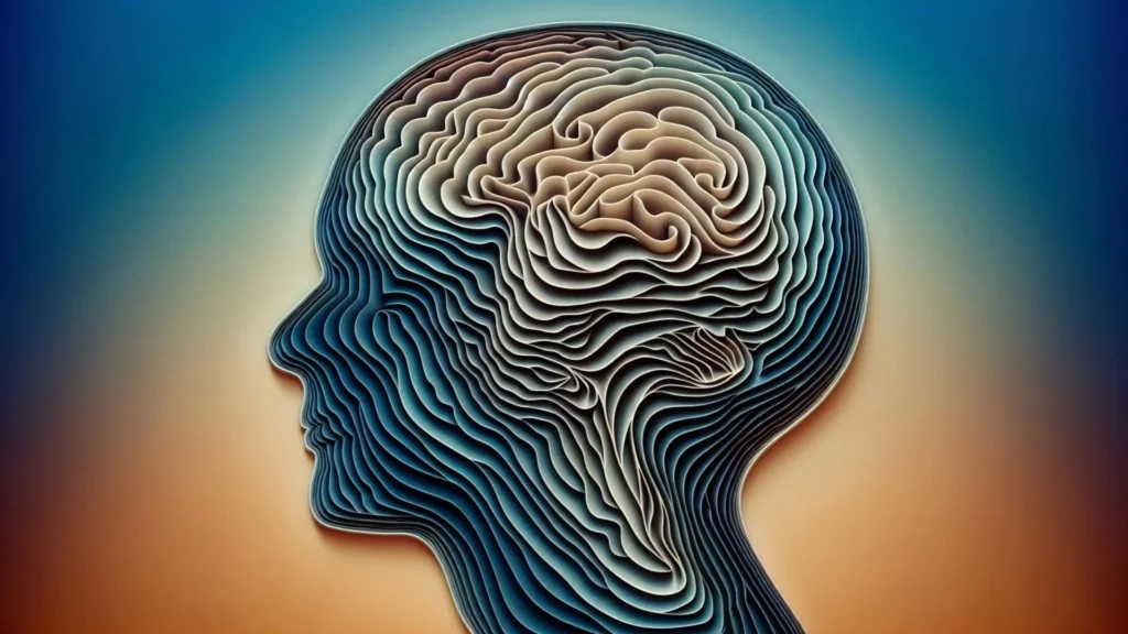
Hidden "Stripes": Unveiling Internal Brain Architecture
Maricarmen Pachicano, a doctoral researcher at the Stevens INI’s Center for Integrative Connectomics and co-first author of the paper, vividly described the visual impact of their findings. "When we visualized gene RNA patterns at single-cell resolution, we could see clear stripes, like geological layers in rock, each representing a distinct neuron type," she stated. This analogy powerfully conveys the distinct, stratified nature of the discovery. "It’s like lifting a veil on the brain’s internal architecture. These hidden layers may explain differences in how hippocampal circuits support learning and memory."
Indeed, the observation that these layers form sheet-like structures that vary in thickness and shape along the hippocampus’s length is particularly significant. This non-uniformity suggests that the functional specialization within CA1 is not merely a global property but rather a finely tuned, regional characteristic. For instance, one section of CA1 might have a more prominent layer associated with spatial memory, while another section might emphasize a layer crucial for emotional memory, thereby explaining the nuanced functional roles attributed to different parts of the hippocampus.
Implications for Neurological Disorders: A New Path to Understanding Vulnerability
The hippocampus is notoriously one of the first brain regions to be affected in the progression of Alzheimer’s disease, manifesting as memory loss, disorientation, and cognitive decline. It also plays a significant role in the pathophysiology of epilepsy, with hippocampal sclerosis being a common finding in temporal lobe epilepsy, and is implicated in other neurological conditions like depression. The identification of CA1’s layered structure provides a critical new framework for understanding why certain neurons are selectively vulnerable in these disorders.
If, for example, a specific neurodegenerative process in Alzheimer’s disease preferentially targets neurons within one of these four layers, the impact on memory and cognition would depend on where along the hippocampus that particular layer is most prominent or active. This offers a more granular understanding than previously possible, moving beyond the general concept of "hippocampal damage" to pinpointing specific cellular populations at risk. This precision could revolutionize diagnostic approaches, allowing for earlier detection of disease-specific neuronal loss, and guide the development of targeted therapeutic interventions aimed at protecting these vulnerable cell types. For epilepsy, understanding which layers contribute to seizure generation and propagation could lead to more effective anti-epileptic drug strategies or surgical targets.
Advancing Brain Mapping with Modern Imaging and Data Science: A Collaborative Future
This discovery stands as a testament to the transformative power of integrating modern imaging technologies with sophisticated data science methodologies. Arthur W. Toga, PhD, director of the Stevens INI and the Ghada Irani Chair in Neuroscience at the Keck School of Medicine of USC, emphasized this point. "Discoveries like this exemplify how modern imaging and data science can transform our view of brain anatomy," Dr. Toga remarked. He highlighted that this work is a continuation of the Stevens INI’s long-standing commitment to mapping the brain at every scale, from molecular interactions to vast neural networks. Such comprehensive mapping efforts are indispensable for both foundational neuroscience research and the translation of discoveries into clinical applications targeting memory and cognition.
The collaborative and open science ethos of the Stevens INI is further exemplified by the team’s commitment to making their findings accessible to the global scientific community. They have compiled their comprehensive data into a new CA1 cell-type atlas, leveraging existing data from the Hippocampus Gene Expression Atlas (HGEA). This invaluable resource is freely available to scientists worldwide, fostering collaboration and accelerating future research. A particularly innovative feature of this atlas is its integration with interactive 3D visualizations, accessible through the Schol-AR augmented-reality app, developed at the Stevens INI. This cutting-edge tool allows researchers to virtually explore the layered structure of the hippocampus in unprecedented detail, manipulating and analyzing the data in an immersive environment.
A Cross-Species Perspective and Future Frontiers
While the current study was conducted using mouse models, the researchers believe that the observed layered pattern may have broad implications across mammalian species. Preliminary observations suggest that this organizational principle, including comparable variations in CA1 thickness, might be conserved in primates and humans. This cross-species similarity provides a strong foundation for future research, prompting investigations into how closely this intricate structure in the human hippocampus matches what has been unveiled in mice. Such comparative studies are crucial for bridging the gap between animal models and human physiology, enhancing the translatability of findings.
The immediate next frontier for this research involves elucidating the functional consequences of this newly discovered architecture. "Understanding how these layers connect to behavior is the next frontier," Bienkowski affirmed. "We now have a framework to study how specific neuron layers contribute to such different functions like memory, navigation, and emotion, and how their disruption may lead to disease." This will involve investigating how each of the four layers participates in specific cognitive tasks, how they interact within the broader hippocampal circuit, and how their individual vulnerabilities manifest in the context of neurological and psychiatric disorders. Such investigations could employ advanced electrophysiological recordings, optogenetic manipulations, and sophisticated behavioral assays to probe the precise roles of these newly defined neuronal populations.
This discovery represents a significant leap forward in neuroscience, moving beyond macroscopic views of brain regions to a molecularly defined, stratified understanding of cellular organization. By peeling back the layers of complexity in the hippocampus, USC researchers have not only redefined a fundamental aspect of brain anatomy but have also illuminated new avenues for understanding, diagnosing, and potentially treating devastating neurological diseases that impact millions globally. The open access atlas and advanced visualization tools further ensure that this foundational work will empower a new generation of researchers to explore the mysteries of memory and cognition.
About the Study and Funding
The study’s impactful findings are the result of a dedicated team effort. In addition to Dr. Michael S. Bienkowski and Maricarmen Pachicano, the other contributing authors include Shrey Mehta, Angela Hurtado, Tyler Ard, Jim Stanis, and Bayla Breningstall.
This pioneering research received crucial financial support from several prestigious institutions, highlighting the collaborative investment in advancing brain science. Funding was provided by the National Institutes of Health/National Institute of Aging (specifically through awards K01AG066847, R36AG087310-01, and a supplement P30-AG066530-03S1), the National Science Foundation (grant 2121164), and generous support from the USC Center for Neuronal Longevity. Furthermore, research data reported in this publication was supported by the Office of the Director, National Institutes of Health under award number S10OD032285, underscoring the broad institutional backing for this critical investigation.