


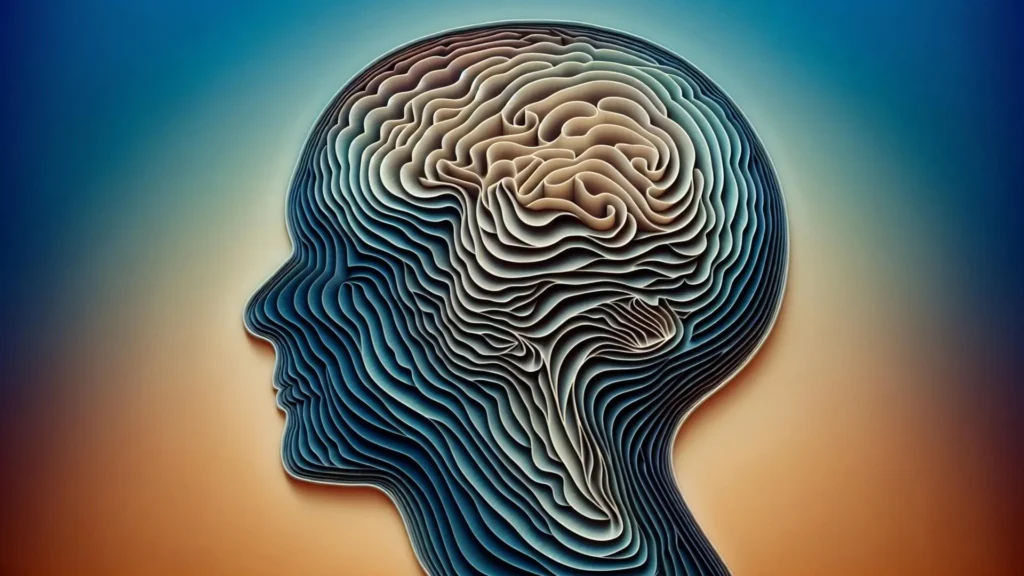
Researchers at the Mark and Mary Stevens Neuroimaging and Informatics Institute (Stevens INI) at the Keck School of Medicine of USC have identified a previously unrecognized, intricate organizational pattern within the CA1 section of the hippocampus, a critical brain region for learning and memory. Published in Nature Communications, their findings reveal that the CA1, long considered a more homogenous or blended area, is actually comprised of four distinct, continuous layers of specialized cell types in mice. This discovery significantly advances the understanding of how information is processed within this vital brain structure, which is fundamental to memory formation, spatial navigation, and emotional regulation. Furthermore, it offers crucial new insights into why certain neuronal populations within the hippocampus are particularly susceptible to neurodegenerative and neurological conditions such as Alzheimer’s disease and epilepsy, paving the way for more targeted research and potential therapeutic interventions.
Deciphering the Brain’s Architectural Blueprint
For decades, the hippocampus has captivated neuroscientists due to its indispensable role in cognitive functions. Damage to this seahorse-shaped structure, famously highlighted by cases like Patient H.M., results in profound amnesia, underscoring its centrality to the formation of new long-term memories. Within the hippocampus, the Cornu Ammonis 1 (CA1) subregion acts as a primary output station, integrating information from other hippocampal subregions (CA3, dentate gyrus) and relaying it to cortical areas. While researchers had long suspected functional specialization within CA1, the precise anatomical and cellular arrangement that underpinned these differences remained elusive. Previous models often depicted CA1 as a somewhat uniform or mosaic mixture of cell types, making it challenging to link specific cellular vulnerabilities to broader neurological dysfunction.
Dr. Michael S. Bienkowski, senior author of the study and an assistant professor of physiology and neuroscience and of biomedical engineering, emphasized this long-standing puzzle. "Researchers have long suspected that different parts of the hippocampus’ CA1 region handle different aspects of learning and memory, but it wasn’t clear how the underlying cells were arranged," he explained. The new study provides a definitive answer, demonstrating that CA1 neurons are not randomly distributed but are instead meticulously organized into four thin, continuous bands. Each band, or layer, represents a distinct neuron type, uniquely defined by a specific molecular signature—a genetic fingerprint that dictates its identity and function. These layers are not static; they exhibit subtle shifts in thickness and position along the length of the hippocampus, implying that each segment of CA1 possesses a unique composition of neuronal types. This nuanced arrangement offers a compelling explanation for the observed functional diversity across the CA1 region, where different segments contribute to distinct behavioral processes. Critically, this differential layering also provides a framework for understanding why certain CA1 neurons might be selectively targeted and made more vulnerable in conditions like Alzheimer’s disease and epilepsy. If a disease process preferentially impacts a specific cell type, its effects would manifest differently depending on where that cell type’s layer is most prominent within CA1.
A Glimpse into Cellular Distinction: The Power of High-Resolution RNA Imaging
The groundbreaking resolution of this discovery was made possible by the application of advanced molecular imaging techniques. The research team employed an RNA labeling technique known as RNAscope, combined with state-of-the-art high-resolution microscopy. This sophisticated approach allowed scientists to visualize single-molecule gene expression directly within mouse CA1 tissue. Unlike traditional staining methods that might only identify broad categories of cells, RNAscope enables the precise identification of individual neuron types based on their unique complement of active genes. Genes, the fundamental units of heredity, are transcribed into RNA molecules, which then serve as blueprints for protein synthesis or perform regulatory functions. By observing which RNA molecules are present and in what quantities within individual cells, researchers can deduce the cell’s specific identity and functional state.
In a meticulous effort, the scientists recorded over 330,000 RNA molecules from 58,065 CA1 pyramidal cells. Pyramidal cells are the principal neurons of the hippocampus, responsible for transmitting information. These recorded RNA molecules represent the genetic instructions that indicate when and where genes are expressed, providing an unprecedented level of detail about the cellular landscape. By systematically mapping these intricate gene activity patterns, the team successfully constructed a detailed cellular atlas. This atlas meticulously outlined the precise boundaries between distinct nerve cell types across the entire CA1 region, transforming the previous understanding of its internal architecture.
The results unequivocally demonstrated that CA1 is composed of four continuous layers of nerve cells, each unequivocally distinguished by its own characteristic pattern of active genes. When reconstructed in three dimensions, these layers form elegant, sheet-like structures that vary in thickness and shape along the longitudinal axis of the hippocampus. This well-defined, stratified arrangement fundamentally revises earlier descriptions that portrayed CA1 as a more blended or mosaic mixture of cell types. The clarity provided by this study resolves long-standing ambiguities and establishes a new anatomical paradigm for the CA1 region.
Hidden "Stripes": Unveiling the Brain’s Internal Architecture
The visual evidence of this discovery was particularly compelling. Maricarmen Pachicano, a doctoral researcher at the Stevens INI’s Center for Integrative Connectomics and co-first author of the paper, vividly described the findings. "When we visualized gene RNA patterns at single-cell resolution, we could see clear stripes, like geological layers in rock, each representing a distinct neuron type," she stated. This geological analogy effectively conveys the distinct and continuous nature of these newly identified layers, suggesting a fundamental and ordered underlying structure that had previously remained obscured. "It’s like lifting a veil on the brain’s internal architecture. These hidden layers may explain differences in how hippocampal circuits support learning and memory." This insight is profound, suggesting that the spatial arrangement of these cell types is directly correlated with their functional contributions to various cognitive processes.
The hippocampus is particularly vulnerable to a range of devastating neurological conditions. It is one of the first brain regions to exhibit damage in Alzheimer’s disease, contributing to the hallmark memory loss associated with the condition. Alzheimer’s disease affects an estimated 6.7 million Americans aged 65 and older, with projections indicating this number could rise to nearly 13 million by 2050. Furthermore, the hippocampus plays a critical role in the generation and propagation of epileptic seizures; epilepsy affects approximately 50 million people worldwide, with a significant portion experiencing drug-resistant forms linked to hippocampal dysfunction. Dysfunction of the hippocampus is also implicated in depression and other psychiatric disorders. The identification of CA1’s layered structure provides an invaluable guide for pinpointing precisely which neuron types may be most at risk as these disorders progress. This specificity is crucial; understanding which cells are affected, and where, is a prerequisite for developing targeted therapies that can protect vulnerable neurons or restore their function. For instance, if a specific cell type in a particular layer is found to be disproportionately affected in early Alzheimer’s, researchers could focus on developing drugs or interventions that specifically target that cell population, potentially halting or slowing disease progression more effectively than broad-spectrum approaches.
Advancing Brain Mapping: The Synergy of Modern Imaging and Data Science
This discovery represents a significant leap in neuroscientific methodology, showcasing the transformative power of integrating advanced imaging techniques with sophisticated data science. Dr. Arthur W. Toga, director of the Stevens INI and the Ghada Irani Chair in Neuroscience at the Keck School of Medicine of USC, underscored this synergy. "Discoveries like this exemplify how modern imaging and data science can transform our view of brain anatomy," he remarked. The Stevens INI has a long and distinguished history in brain mapping, operating at every conceivable scale, from the molecular intricacies of individual cells to the complex connectivity of whole brain networks. This latest work not only adds a new layer of detail to the institute’s extensive atlas of brain architecture but also promises to inform both fundamental neuroscience investigations into how the brain works and translational studies aimed at treating disorders affecting memory and cognition.
The sheer volume of data generated in modern neuroscience studies necessitates robust computational tools for analysis and visualization. The ability to process hundreds of thousands of individual RNA molecules from tens of thousands of cells, and then reconstruct their spatial organization in three dimensions, is a testament to the advancements in bioinformatics and data science. These tools enable researchers to identify subtle patterns and relationships that would be impossible to discern through traditional manual examination, pushing the boundaries of what can be discovered about brain structure and function. The collaboration between imaging specialists, molecular biologists, and data scientists is increasingly vital in tackling the immense complexity of the brain. Such interdisciplinary approaches are driving a new era of "big data" neuroscience, where comprehensive datasets are curated and analyzed to reveal previously hidden biological principles.
A New CA1 Cell-Type Atlas: A Global Resource for Research
In a commitment to open science and collaborative research, the team has compiled its extensive findings into a new CA1 cell-type atlas. This invaluable resource is built upon data from the Hippocampus Gene Expression Atlas (HGEA) and is freely available to scientists worldwide. The atlas includes interactive 3D visualizations, made accessible through the Schol-AR augmented-reality app, a cutting-edge tool developed at the Stevens INI. This innovative app allows researchers globally to explore the newly defined layered structure of the hippocampus in unprecedented detail, facilitating deeper understanding and inspiring new hypotheses. By making this data publicly available, the USC team aims to accelerate the pace of discovery across the global neuroscience community, enabling other laboratories to build upon their foundational work without proprietary barriers. This sharing of resources is crucial for collaborative progress in complex fields like brain research, fostering a more inclusive and efficient scientific ecosystem.
The potential for broad applicability of these findings is further bolstered by comparative anatomical observations. The layered pattern identified in mice exhibits remarkable resemblance to similar arrangements observed in primates and humans, including comparable variations in CA1 thickness. This suggests that the fundamental organizational principles of the hippocampus, at least within the CA1 subregion, may be conserved across a wide range of mammalian species. While further research is explicitly needed to determine the exact degree of structural homology between the mouse model and the human brain, these findings provide a robust starting point. They establish a strong framework for future studies that will meticulously examine how hippocampal architecture, particularly this newly discovered layered organization, underpins memory, spatial cognition, and emotional processing in humans. The use of mouse models in neuroscience is a long-standing tradition, providing crucial insights into basic biological mechanisms that are often conserved across species, accelerating the path toward understanding human brain function and disease.
Implications for Disease and the Future of Neuroscience
The implications of this discovery are far-reaching, offering new avenues for understanding and potentially treating devastating neurological conditions. Alzheimer’s disease, affecting millions globally, is characterized by progressive memory loss and cognitive decline, with the hippocampus being an early and significant site of pathology. The ability to identify specific vulnerable cell types within these newly defined layers could lead to the development of highly targeted therapies. Instead of general neuroprotective agents, future treatments might focus on preserving or regenerating neurons in a particular CA1 layer that is most susceptible to early amyloid-beta plaque accumulation or tau pathology. Similarly, in epilepsy, hippocampal sclerosis (scarring of the hippocampus) is a common feature in drug-resistant temporal lobe epilepsy. Understanding which neuronal layers are involved in seizure generation and propagation could lead to more precise surgical interventions or novel anti-epileptic drugs that modulate the activity of specific cell populations. These targeted approaches represent a significant shift from broad-spectrum treatments, promising greater efficacy and fewer side effects.
Beyond disease, this new understanding of CA1’s architecture has profound implications for basic neuroscience. It provides a more refined structural context for investigating how the hippocampus encodes, stores, and retrieves memories. Researchers can now design experiments to probe the specific contributions of each layer to different aspects of memory, such as episodic memory (memory for events) versus spatial memory (memory for locations). For example, using techniques like optogenetics or pharmacogenetics, scientists could selectively activate or inhibit neurons in a particular layer and observe the resulting effects on an animal’s behavior and cognitive performance. This level of precision was previously unimaginable when CA1 was viewed as a more undifferentiated region. This refined anatomical map will undoubtedly guide functional studies, allowing for a more nuanced interpretation of neuronal activity and connectivity within the hippocampus.
Dr. Bienkowski articulated the next frontier of this research: "Understanding how these layers connect to behavior is the next frontier. We now have a framework to study how specific neuron layers contribute to such different functions like memory, navigation, and emotion, and how their disruption may lead to disease." This signifies a paradigm shift, moving from a general understanding of hippocampal function to a granular, layer-specific analysis. The development of advanced computational models incorporating this new architectural detail will also be crucial for simulating hippocampal circuit dynamics with greater accuracy, providing theoretical frameworks to test experimental findings. The potential for this discovery to accelerate research into complex cognitive processes and neurological disorders cannot be overstated.
The study, which also included authors Shrey Mehta, Angela Hurtado, Tyler Ard, Jim Stanis, and Bayla Breningstall, was supported by significant funding from the National Institutes of Health/National Institute of Aging (K01AG066847, R36AG087310-01, supplement P30-AG066530-03S1), the National Science Foundation (grant 2121164), and the USC Center for Neuronal Longevity. This robust support underscores the recognized importance of fundamental research into brain architecture and its direct relevance to human health. The research data reported in this publication was also supported by the Office of the Director, National Institutes of Health under award number S10OD032285, highlighting the collaborative and multi-institutional nature of cutting-edge scientific endeavors. As neuroimaging and molecular biology continue to evolve, discoveries like this promise to continually refine our "atlas" of the brain, bringing us closer to unlocking its deepest secrets and developing effective treatments for its most debilitating disorders.