





The landscape of aerospace medicine underwent a fundamental transformation following the successful deployment and operation of a portable X-ray system during the SpaceX Fram2 mission. For the first time in over forty years, the monopoly of ultrasound as the sole diagnostic imaging modality in space has been challenged, opening new frontiers for astronaut health and mission safety. This milestone, detailed in a study published in the journal Radiology, demonstrates that high-quality diagnostic imaging can be achieved in microgravity by crew members with minimal medical training, utilizing commercial off-the-shelf technology.
The Four-Decade Dominance of Spacebound Ultrasound
Since the early 1980s, ultrasound has been the "gold standard" and, effectively, the only standard for medical imaging in the extraterrestrial environment. NASA and other international space agencies favored ultrasound due to its lack of ionizing radiation, its relatively compact size, and its ability to visualize soft tissue. However, ultrasound is notoriously operator-dependent. On the International Space Station (ISS), astronauts often require extensive, specialized training or real-time remote guidance from Earth-based sonographers to capture usable images.
Furthermore, the physical environment of a spacecraft presents unique challenges for soundwave-based imaging. The interior of a spacecraft is a cacophony of mechanical hums, fan whirs, and life-support systems, creating a "loud" acoustic environment that can interfere with sensitive equipment. Moreover, ultrasound is primarily effective for soft tissue, such as checking for kidney stones or monitoring fluid shifts in the eye—a condition known as Space-Associated Neuro-ocular Syndrome (SANS). It is significantly less effective for diagnosing bone fractures or internal structural failures in hardware, areas where X-ray technology excels.
Technical Barriers to Extraterrestrial Radiography
The transition from ultrasound to X-ray was long delayed by three primary factors: mass, power, and stability. Traditional X-ray machines found in hospitals are massive, immobile installations that require heavy lead shielding to protect both the operator and the surrounding environment from radiation. In the weight-sensitive world of rocketry, where every kilogram costs thousands of dollars to launch, a standard X-ray suite was a logistical impossibility.
Radiation itself posed another concern. Space is already a high-radiation environment; introducing a device that generates additional ionizing radiation required careful calibration to ensure the cumulative dose to the crew remained within safety limits. Finally, the "conceit" of space medicine, as noted by Mayo Clinic researcher Sheyna Gifford, was that the constant motion of microgravity would make it impossible for a subject to remain still enough for a clear diagnostic exposure. On Earth, patients are told to "hold their breath" and stay perfectly still; in orbit, subjects and the machine itself are in a state of perpetual freefall.
The Path to the Fram2 Breakthrough: A Chronology
The success of the 2025 Fram2 mission was the culmination of years of iterative testing and collaboration between the Mayo Clinic, SpaceX, and MinXray Inc. The timeline of this breakthrough began in earnest in 2022, when Dr. Sheyna Gifford and her team conducted a series of feasibility studies during parabolic flights. These "vomit comet" missions provided brief 20-second bursts of microgravity, allowing researchers to prove that digital X-ray detectors could capture images without blurring despite the lack of gravity.
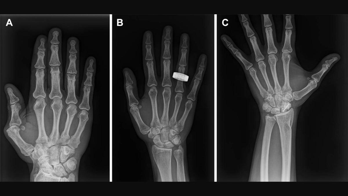
Following the success of the parabolic trials, the team spent two years refining the imaging protocols and hardware. They selected the MinXray TR90BH, a portable X-ray generator roughly the size of an ice cooler, known for its use in remote field medicine and veterinary practice on Earth. The goal was to prove that this commercial technology could withstand the rigors of a rocket launch and remain functional in the vacuum-like isolation of orbit.
On March 31, 2025, the SpaceX Falcon 9 rocket launched the Fram2 mission into a polar orbit. The crew, consisting of three non-medical professionals, carried the portable radiography kit as a primary research payload. Prior to launch, the crew underwent a condensed training program lasting only four hours—a stark contrast to the weeks or months of training usually required for complex medical procedures in space.
Experimental Methodology and Data Collection
The experimental protocol was designed to be rigorous and comparative. Before departing Earth, the crew used the MinXray device to take baseline radiographs of several body parts: the hand, forearm, chest, abdomen, and pelvis. These "preflight" images served as the control group.
Once the Dragon spacecraft reached stable orbit, the crew initiated the "in-flight" phase. On the first day of the mission (L+1), radiographs of the hand were acquired. By the third day (L+3), the crew performed more complex scans of the chest and abdomen. To test the versatility of the machine beyond human biology, they also performed an X-ray of a smartwatch, demonstrating the potential for non-destructive testing (NDT) of mission-critical electronics.
Upon the mission’s conclusion and the crew’s return to Earth, a "postflight" set of images was taken by professional radiographers using the same equipment and protocols. This created a three-tiered dataset (preflight, in-flight, and postflight) that allowed researchers to isolate the effects of microgravity on image quality.
Analyzing the Results: Clinical Quality in Orbit
The images were subjected to a "blind" review by three independent radiologists. These experts evaluated the scans based on several clinical criteria, including positioning accuracy, spatial resolution, contrast resolution, and overall diagnostic utility.
The results were overwhelmingly positive. The radiologists concluded that the in-flight images of the extremities (hand and forearm) were virtually indistinguishable from those taken on Earth. While the positioning scores for the central body images (chest and pelvis) showed a slight decrease—likely due to the difficulty of tethering a floating body in a cramped capsule—the spatial and contrast resolutions remained high. Every scan produced in orbit was deemed "diagnostically useful," meaning a doctor could use them to make a definitive medical diagnosis.

The crew’s feedback was equally vital. Despite the minimal four-hour training window, the astronauts reported that the machine was intuitive and easy to operate. This suggests that in a future emergency scenario—such as a suspected rib fracture or a lung complication—a layperson astronaut could provide life-saving diagnostic data to flight surgeons on Earth.
Implications for Deep Space Exploration and Infrastructure
The success of the Fram2 X-ray experiment has implications that extend far beyond the immediate health of the crew. As humanity looks toward sustained lunar bases and multi-year missions to Mars, the ability to "see through" objects becomes a necessity for survival.
Dr. Gifford highlighted that X-rays are critical for maintaining the integrity of the mission’s hardware. In deep space, an astronaut’s spacesuit is their only protection against a lethal environment. If a suit is nicked by a micrometeoroid or experiences a mechanical failure in its life-support backpack, an X-ray can inspect the internal components without the risk of disassembling the pressurized suit. Similarly, as electronics are exposed to cosmic radiation over long durations, internal solder joints can fail or "whiskers" can grow on circuits; portable radiography allows for the inspection of these delicate systems in situ.
Furthermore, the democratization of medical imaging is a key takeaway. The move toward "autonomous medicine"—where the crew can diagnose and treat themselves without heavy reliance on Earth-based experts—is essential for Mars missions, where communication delays can reach up to 20 minutes each way.
Future Challenges and Technical Refinements
Despite the success of the mission, the study also identified areas for improvement. The Fram2 crew reported that the MinXray unit sustained minor exterior damage during the high-G forces of launch and the jarring impact of splashdown. While the internal components remained functional, future iterations of space-rated X-ray machines will require enhanced ruggedization to survive the vibrations of next-generation heavy-lift rockets like SpaceX’s Starship.
Additionally, researchers are looking to further reduce the size and power requirements of the units. Future models may incorporate carbon nanotube (CNT) X-ray emitters, which are lighter and more energy-efficient than traditional vacuum tubes.
The Fram2 mission has effectively closed the door on the era of "ultrasound-only" space medicine. By proving that portable, commercial X-ray technology can deliver clinical-grade results in the harshest of environments, the study paves the way for a more robust and resilient healthcare infrastructure for the next generation of space explorers. As missions become longer and move further from Earth, the ice-cooler-sized box that once seemed a "dream" for aerospace medicine is now a proven reality.